Danger: White Spots on Your Skin and the "Fatal" Mistakes in Treatment
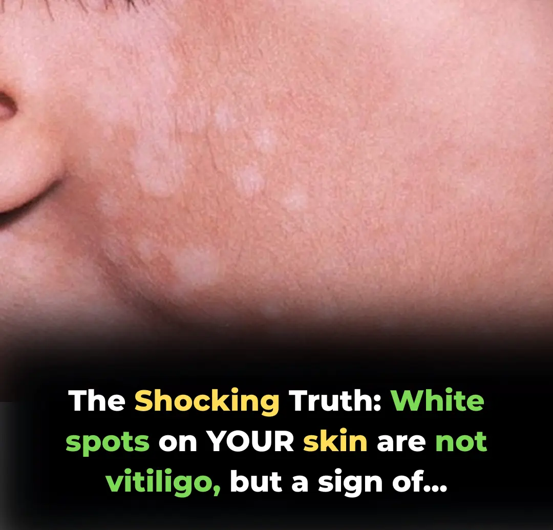
Most people panic the moment they see a white patch on their skin, immediately Googling "Vitiligo" and fearing the worst. However, in the world of dermatology, not every white spot is a life-long autoimmune struggle. In fact, misdiagnosing yourself and applying the wrong treatment can lead to permanent skin damage, scarring, or chronic fungal infections.
This guide will break down the four most common culprits of skin depigmentation, providing expert-level insights into their causes, symptoms, and clinically-backed solutions.
1. Tinea Versicolor (The Fungal Imposter)
This is the most frequent cause of white spots, especially in tropical or high-humidity climates.
The Science Behind the Spots
Tinea Versicolor is caused by an overgrowth of Malassezia, a yeast-like fungus naturally present on human skin. When it grows out of control, it secretes Azelaic Acid. This specific acid inhibits the production of melanin (your skin's natural pigment), leaving behind pale "ghost" spots.
Clinical Manifestations
-
Appearance: Well-defined macules that can be white, pink, or light brown.
-
Texture: Often covered in a fine, "furfuraceous" (flour-like) scale.
-
Sensation: Mild pruritus (itching) that intensifies when the body temperature rises or during perspiration.
Expert-Approved Solutions
-
Topical Antifungals: Shampoos or body washes containing 2% Ketoconazole or Selenium Sulfide should be applied to the skin for 5-10 minutes before rinsing.
-
Azole Creams: For localized spots, creams like Clotrimazole or Terbinafine are highly effective.
-
Environmental Control: Wear breathable cotton fabrics and keep the skin as dry as possible to prevent fungal recurrence.
2. Pityriasis Alba (The "Dry Skin" Echo)
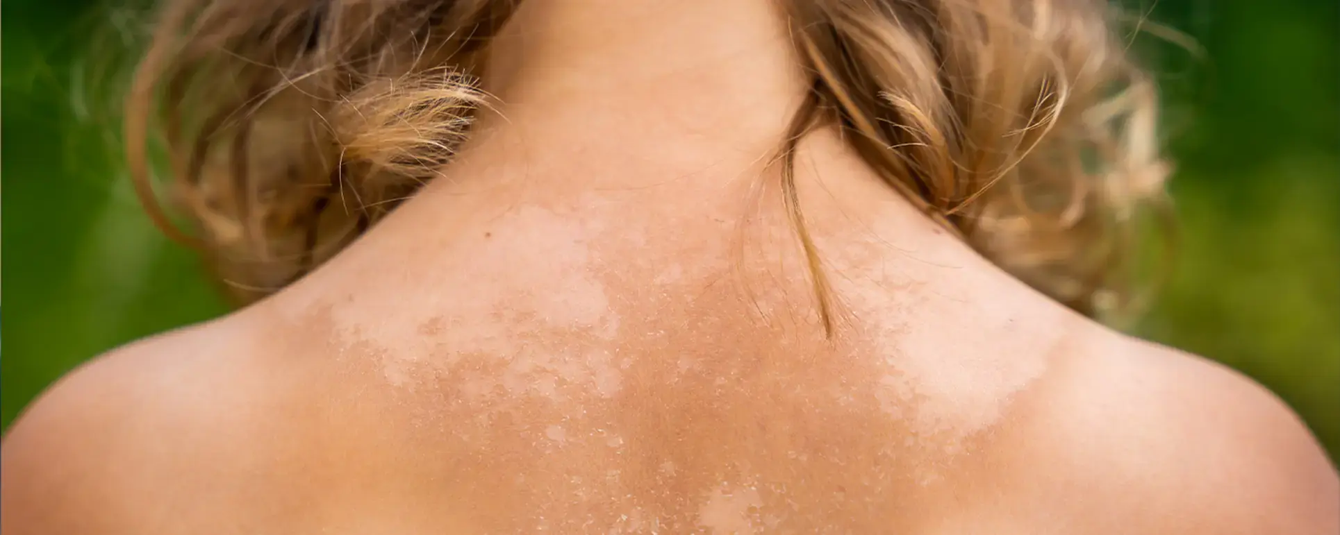
Often mistaken for fungus or "worm-ring" spots, Pityriasis Alba is actually a mild, localized form of Atopic Dermatitis (Eczema).
The Root Cause
While the exact etiology remains debated, it is closely linked to Xerosis (extreme skin dryness) and excessive, unprotected sun exposure. It is most commonly seen in children and adolescents.
Visual Indicators
-
Morphology: Round or oval patches, typically 0.5 cm to 2 cm in diameter.
-
Evolution: It begins as a slightly red, scaly patch that eventually fades into a pale, hypopigmented spot.
-
Location: Primarily targets the cheeks, forehead, and upper arms.
Therapeutic Approach
-
Intensive Rehydration: The cornerstone of treatment is using fragrance-free, hypoallergenic emollients (moisturizers) multiple times a day.
-
Photoprotection: Broad-spectrum sunscreen is mandatory. By preventing the surrounding skin from tanning, the white spots become significantly less noticeable.
-
Gentle Cleansing: Avoid harsh alkaline soaps that strip the skin’s natural lipid barrier.
3. Idiopathic Guttate Hypomelanosis (The "White Freckles")
Think of these as the "reverse" of age spots. They are essentially white freckles caused by long-term skin maturation.
The Biological Mechanism
Years of cumulative Ultraviolet (UV) damage cause the melanocytes (pigment cells) in specific spots to simply "turn off" or die out. This condition is most prevalent in individuals over the age of 40.
Identifying the Signs
-
Features: Small, porcelain-white, "drop-like" spots (2-5 mm).
-
Texture: Smooth and slightly thinned (atrophic) skin surface.
-
Distribution: Almost exclusively on sun-exposed areas like the shins and forearms.
Management and Prevention
-
Retinoids: Prescription-strength topical retinoids can stimulate cell turnover and may help partially restore pigment.
-
Advanced Procedures: Cryotherapy (liquid nitrogen) or fractional laser resurfacing can be used by specialists to "reboot" the skin's pigment production.
-
Rigorous Shielding: Once these spots appear, they are difficult to reverse; preventing new ones via UPF-rated clothing is essential.
4. Vitiligo (The Autoimmune Challenge)
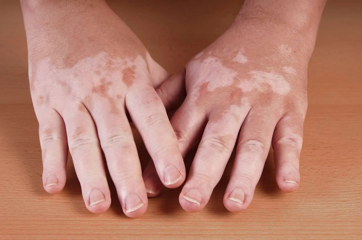
Unlike the conditions above, Vitiligo is a systemic autoimmune disorder and requires a completely different medical strategy.
The Underlying Pathogenesis
The body’s own immune system mistakenly identifies melanocytes as foreign invaders and destroys them. This results in a total loss of pigment in the affected areas.
Distinctive Symptoms
-
Color: A stark, "chalk-white" or "milky-white" appearance.
-
Borders: Extremely sharp, distinct margins.
-
Leukotrichia: Hair growing within the white patch may also turn white.
-
Symmetry: Often appears symmetrically on both sides of the body (e.g., both knees or both hands).
Specialized Treatments
-
Phototherapy: Narrowband UVB (NB-UVB) is the gold standard for stimulating repigmentation.
-
Immunomodulators: Topical corticosteroids or Calcineurin inhibitors (Tacrolimus) to calm the immune response.
-
Surgical Options: In stable cases, melanocyte-keratinocyte transplant or skin grafting may be considered.
The "Fatal" Mistakes: What NOT To Do
-
Self-Medicating with Steroids: Applying strong steroid creams to Tinea Versicolor (fungus) will act as "fuel," causing the infection to spread rapidly while thinning your skin.
-
Aggressive Scrubbing: Trying to "scrub off" white spots often leads to Post-Inflammatory Hyperpigmentation or scarring, making the patches even more visible.
-
Ignoring the Sun: Assuming that "tanning" will hide white spots. In reality, tanning darkens the surrounding skin, making the white patches stand out like neon signs.
Final Verdict
If your white spots are spreading rapidly, losing their borders, or affecting your hair color, skip the home remedies and consult a board-certified dermatologist immediately.